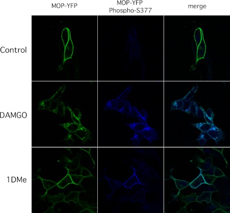
FIGURE 5.
Differential localization of Ser-377-phosphorylated MOP receptors after DAMGO or 1DMe treatment in (SH2-D9)MOP-YFP cells. Cells were treated with buffer alone, 1 μm DAMGO, or 1DMe for 30 or 15 min at room temperature, respectively, before formaldehyde fixation. Total MOP receptors were visualized using the fluorescence of the YFP tag. MOP receptors phosphorylated on Ser-377 were visualized by immunofluorescence using the phospho-specific MOP antibody. Confocal images were taken with a Zeiss 710 NLO inverted microscope with ×40 objective as described under “Experimental Procedures.”

