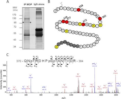
FIGURE 7.
NanoLC-MS/MS analysis of MOP receptor phosphorylation sites. A, immunoprecipitated (IP) MOP receptors visualized after colloidal Coomassie staining (between asterisks). IgG alone correspond to the antibody used for immunoprecipitation, without cellular extract. MW, molecular weight. B, amino acid sequence of the part of the C-tail of MOP that was covered by our study. The putatively palmitoylated cysteine is shown. Phosphorylated residues are shown in red. Trypsin cleavage sites are in yellow. The gray sequence corresponds to the beginning of the YFP fusion. C, ETD MS/MS spectrum of the triply phosphorylated peptide, 370QNpTRDHPpSpTANTVDR384 (triply charged precursor ion, MH3+, at m/z 651.2417), displays series of c- and z-ions indicating that Thr-372, Ser-377, and Thr-378 are phosphorylated. Sequence and phosphorylated positions are indicated.

