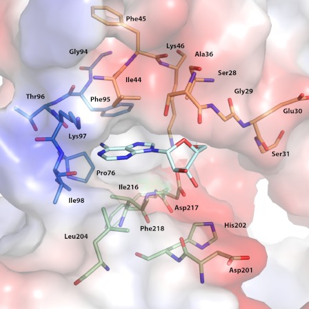
FIGURE 1.
Key components of nucleoside-binding site. Residues forming the nucleoside-binding site of APH(2″)-IVa are shown. A bound adenosine molecule is shown in cyan stick representation. Key residues are divided into three regions based on secondary structure elements and color-coded as follows: the N-terminal β-strands forming the top face of the cleft are shown in orange, the linker loop is shown in blue, and the loops from the core subdomain forming the bottom face of the cleft are shown in green.

