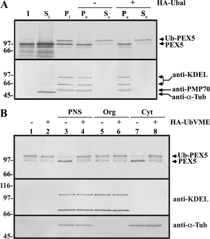
FIGURE 1.
DUB acting on Ub-PEX5 is a cytosolic protein. A, 35S-labeled PEX5 was incubated with a PNS in the presence of AMP-PNP for 20 min at 37 °C. The import reaction was then separated into soluble (Si) and organelle (Pi) components by centrifugation. The organelles were resuspended in ATP-containing buffer, in the absence or presence of HA-Ubal (lanes − and +, respectively), and further incubated for 20 min at 37 °C. Finally, the suspensions were separated into an organelle pellet (Pe) and a supernatant (Se) by centrifugation. Samples were treated with 20 mm N-ethylmaleimide and subjected to SDS-PAGE under nonreducing conditions followed by Western blot. The membrane was first exposed to an x-ray film to detect the 35S-labeled PEX5 (top panel) and afterward was sequentially probed with the following antisera: anti-KDEL (recognizes GRP72 and GRP98, two endoplasmic reticulum proteins); anti-α-tubulin (a cytosolic marker; anti-α-Tub), and anti-PMP70 (an intrinsic protein of the peroxisomal membrane). B, 35S-labeled Ub-PEX5 was incubated alone (lanes 1 and 2), with a PNS (lanes 3 and 4), or with the corresponding organelle (Org; lanes 5 and 6) or cytosolic (Cyt; lanes 7 and 8) fractions, in the absence (−) or presence (+) of HA-UbVME, as indicated. Samples were analyzed as above. The distributions of microsomal (anti-KDEL), and soluble proteins (anti-α-Tub) are also shown. Lane I, 50% of the 35S-labeled PEX5 reticulocyte lysate used in this experiment. Numbers to the left indicate the molecular masses of the reduced protein standards in kDa.

