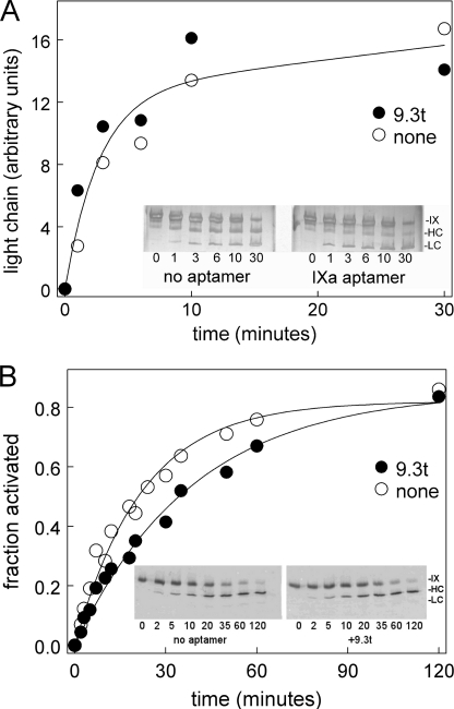
FIGURE 3.
Aptamer effect on factor IX activation. A, factor IX was incubated with the aptamer (●) or a buffer control (○) before being activated with factor XIa in buffer without albumin. At the indicated times, samples were run on a 10–15% gradient PhastGel and silver-stained. Images were analyzed using ImageJ. The band intensity in the light chain (units of the band intensity from ImageJ scans) is plotted versus time. The insets show grayscale images of the gels. B, factor IX was incubated with the aptamer (●) or a buffer control (○) before being activated with factor VIIa-tissue factor. At the indicated times, samples were run on a 10–15% gradient PhastGel and transferred to a nylon membrane for Western blotting with anti-factor IX primary and fluorescent secondary antibodies. Fluorescence was detected with a LI-COR ODYSSEY system. Separate experiments examined reduced and unreduced samples, and all data are shown. The insets show representative Western blots.

