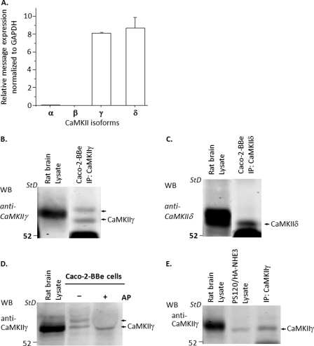
FIGURE 5.
Expression of CaMKII isoforms γ and δ in Caco-2BBe cells and PS120 cells. A, Caco-2BBe cells. Quantitative RT-PCR revealed that messages for only two CaMKII isoforms, CaMKIIγ and CaMKIIδ, were expressed in the Caco-2BBe cells, and they were expressed in similar quantity. B and C, an immunoprecipitation (IP)/Western blot (WB) approach was used with specific anti-CaMKIIγ and anti-CaMKIIδ antibodies to confirm the presence of these two CaMKII isoforms. B, CaMKIIγ (n = 7); C, CaMKIIδ (n = 2) in Caco-2BBe cells. Rat brain was used as a positive control. D, Western blotting of Caco-2BBe cells with CaMKIIγ antibodies reveals two bands. The upper band disappeared after the Caco-2 cell lysate was treated with alkaline phosphatase at 37 °C for 60 min, but there was no change in the lower band (n = 2). E, immunoprecipitation/Western blot was used to identify isoforms of CaMKII proteins expressed in PS120 cells. Only CaMKIIγ was present (n = 5). Specific CaMKIIα and CaMKIIδ antibodies failed to identify these isoforms (data not shown).

