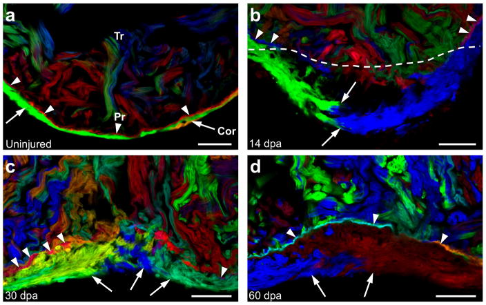
Figure 4. Regeneration of cortical and primordial muscle after injury.
a, Section of an uninjured ventricular apex, indicating the primordial (arrowheads) and cortical (arrows) layers. b, Regenerating ventricular apex at 14 days after resection (dpa). Cortical muscle clones converge within the injury site, while the primordial layer lags behind. Dashed line indicates amputation plane. c, 30 dpa ventricular apex, indicating multiple cortical clones and an incomplete primordial layer. d, Regenerated ventricular apex at 60 dpa, containing cortical muscle overlying a mostly contiguous layer of primordial muscle. n=6 animals for each timepoint. Scale bars, 50 μm

