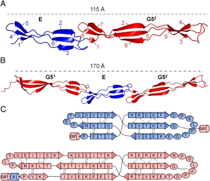
Fig. 3.
Crystal structures of SasG domains; E and G5 domains are shown in blue and red, respectively. (A) Structure of E-G52. The β-strands are numbered for E and G5 domains. (B) Structure of G51-E-G52. (C) Schematic of the secondary structure of E (Upper) and G52 (Lower), as defined by the method of Kabsch and Sanders (64).

