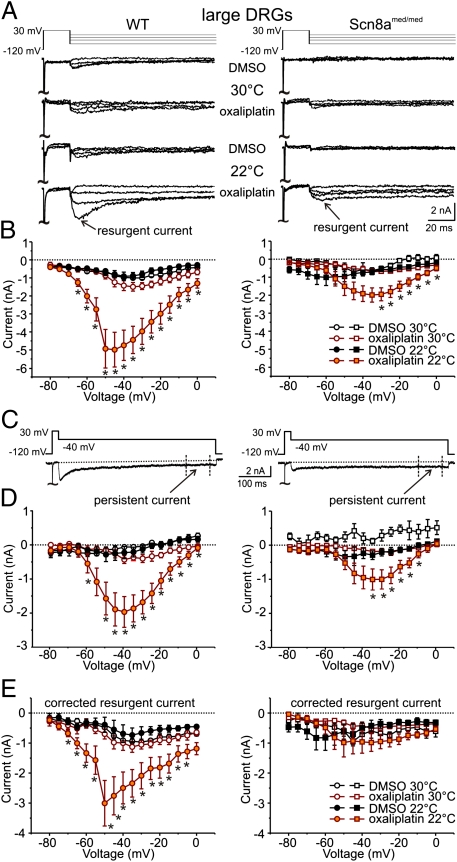
Fig. 4.
Oxaliplatin and cooling enhance INaR and INaP TTX-s Nav currents in large-diameter DRG neurons. (A) Representative current traces in response to voltage commands to −75, −45, −25, and −5 mV (Upper lane) from large-diameter (40.6 ± 4.5 μm, n = 25) DRG neurons from wild-type (WT, Left) and Scn8amed/med mice (Right) at 30 °C and 22 °C after incubation with oxaliplatin (30 μM; ∼90 min). (B) Peak INaR as a function of voltage for recordings at 30 °C [open symbols: WT n = 5;5 and Scn8amed/med: n = 4;6 (DMSO; oxaliplatin)] and 22 °C (filled symbols, WT: n = 10;11, Scn8amed/med: n = 6;8) from DRG neurons incubated with vehicle (1% vol/vol DMSO) or oxaliplatin. (C) Representative voltage command (Upper) and current (Lower) traces indicating the period over which the INaP component was determined (400–475 ms). (D) INaP shown as a function of voltage for recordings at 30 °C (open symbols, WT n = 5;5 and Scn8amed/med n = 8;11 for DMSO; oxaliplatin) and 22 °C (filled symbols, WT: n = 10;11, Scn8amed/med: n = 6;8) from large-diameter DRG neurons (WT Left, Scn8amed/med Right) after incubation with vehicle (black) or oxaliplatin (orange). (E) Corrected INaR amplitude as a function of voltage for recordings at 30 °C (open) and 22 °C (filled) from large-diameter DRG neurons (WT Left, Scn8amed/med Right) after incubation with vehicle (black) or oxaliplatin (orange). INaR was corrected for each individual trace by subtracting the INaP component (D) from the peak INaR (B). Mice ranging in age from P14–25 were used. (*P < 0.05, **P < 0.01).

