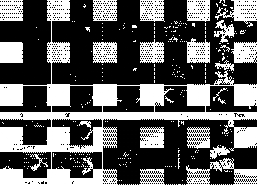
Fig. 3.
Enhancement of protein expression levels by 3′-UTR elements. (A–E) Increase in the level of cytoplasmic GFP generated from 10XUAS constructs by the addition of various 5′- or 3′-UTR elements was determined by measuring native GFP fluorescence using the photomultiplier tube (PMT) of a Zeiss 510 confocal microscope. Dissected nervous systems were imaged under identical conditions except for the Inset in A, which was imaged at higher gain. (A) Level of expression obtained with pJFRC13 (Fig. 1) (29), which was used as a baseline to measure the level of enhancement produce by addition of the other sequence elements. (B) Increase of 6.1-fold in cell body GFP fluorescence was observed when the WPRE element was added to the 3′-UTR in pJFRC14 (29). (C) Increase of 7.5-fold was observed by the addition of the Syn21 element to the 5′-UTR (pJFRC80). (D) p10 element produces a 23-fold increase when added to the 3′-UTR (pJFRC28). (E) Further increases in expression are observed when both the Syn21 and p10 elements are present (pJFRC81). (F–J) Pairs of neurons, viewed transversely, from the same series of genotypes, following staining with anti-GFP antibody. (K and L) Specimens imaged as in F–J using vectors that contain the same UTR elements as pJFRC13 but that express membrane-targeted GFP, either mCD8::GFP (K; pJFRC2; 29) or myr::GFP (L; pJFRC12; 29). M (pJFRC12) and N (pJFRC29) show that the p10 element can also increase GFP expression in the female germline. (O and P) Antibody staining for GFP in lines carrying a 20XUAS-Syn21-Shibire[ts1]::GFP-p10 construct (pJFRC101) inserted at the VK00005 (O) or attP2 (P) genomic integration sites. The R66A12-GAL4 driver was used to generate the data shown in A–L and O and P. The R34C10-GAL4 driver was used for M and N.

