Fig. 2.
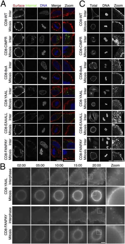
CD8 chimeras endowed with internalization motifs are predominantly localized to the plasma membrane and are not internalized during mitosis. (A) Representative confocal images of anti-CD8 antibody uptake experiments. Uptake was performed as described in Fig. 1A. Internalized CD8 is shown in green, and surface CD8 is shown in red. 3D reconstructions of these cells are shown in Fig. S1. (B) Live cell imaging of Alexa488-conjugated anti-CD8 uptake in cells expressing CD8-YAAL or CD-FANPAY. Note the extensive membrane labeling in mitotic cells. (C) Representative confocal images of the subcellular distribution of CD8 constructs in permeabilized HeLa cells at interphase or mitosis. Each row of images shows anti-CD8 and DAPI staining of permeabilized interphase and mitotic cells expressing the indicated chimeras. (Scale bars: 10 μm.)

