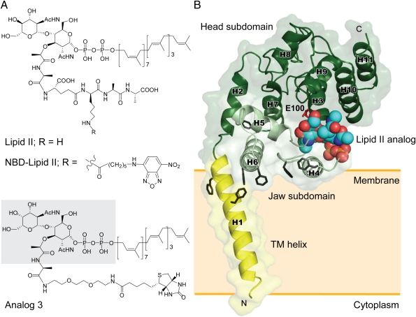
Fig. 1.
Structures of lipid II, lipid II analogs, and SaMGT-analog complex. (A) Chemical structures of lipid II and lipid II analogs. The portion of analog 3 that can be observed on the electron density map is shaded in gray. (B) An overall structure of SaMGT-analog. The TM helix, jaw subdomain, and head subdomain are color-coded in yellow, light green, and dark green, respectively. The lipid II analog 3 is shown as van der Waals spheres. Putative active site, E100 of SaMGT, and aromatic residues are located near the water–membrane interfaces, which are shown as sticks in red and black, respectively. The numbering of helices of SaMGT is indicated as H1–H11. The proposed membrane location is indicated by an orange shaded rectangle. The dissociation constant of analog 3 is ∼12.9 μM.

