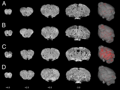
Fig. 2.
MRI detection of VCAM–MPIO binding in 4T1 model. Selected T2*-weighted images from a 3D dataset (coordinates relative to Bregma) at days 5 (A), 10 (B), and 13 (C) after intracardiac injection of 4T1 cells. Intense focal hypointense areas (black) correspond to MPIO binding. No specific MPIO binding was detected in naïve BALB/c mice injected with VCAM–MPIO (D). 3D reconstructions (column 5) show the spatial distribution of VCAM–MPIO binding (red) throughout the brain.

