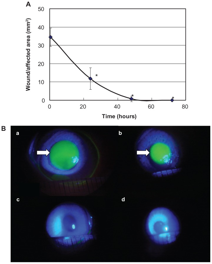
Figure 2.
Evaluation of the wound/affected area method (A) and fluorescein staining (B).
Notes: During the period from 30 minutes to 48 hours after detachment, the wound/affected areas decreased approximately linearly with time. Data are means ± standard deviation of values from four eyes. *P < 0.05 versus the corresponding value for after 30 minutes (Student’s t-test). The wound/affected area as determined by fluorescein staining decreased gradually over time, and there was little chromatic response at 48 hours. The arrow shows the corneal epithelium defect observed by fluorescein staining. a: 30 minutes after detachment; b: 24 hours after detachment; c: 48 hours after detachment; and d: 72 hours after detachment.

