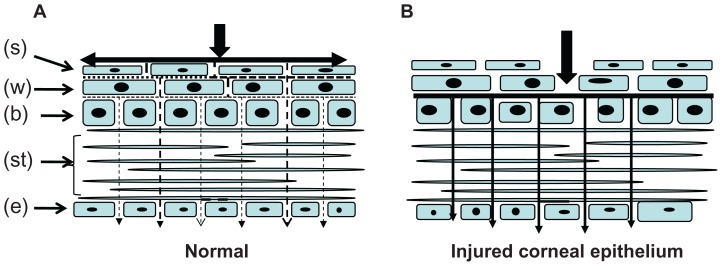
Figure 6.
Microelectrode profiles across the rabbit corneal epithelium.
Notes: A diagram of the cornea showing the epithelium, a portion of stroma (st), endothelium (e), superficial (s) cells, basal (b) cells, and wing (w) cells. (A) The electric current flow is strong in the cornea surface and weak inside the cornea of normal rabbit eyes. (B) The electric current flow to the cornea increased after damage to the cornea. Arrow shows the electric current flow.

