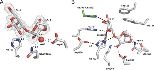
FIGURE 3.
Structural features of neoagarobiose recognition in BpGH117. A, neoagarobiose trapped in the active site of the E303Q mutant of BpGH117. The electron density of the neoagarobiose is shown in gray mesh as a maximum likelihood/σa-weighted Fo − Fc map contoured at 3σ (0.19e/Å3). The three putative catalytic residues are shown in stick representation. B, residues forming hydrogen bonds and hydrophobic interactions with neoagarobiose are shown in stick representations. Hydrogen bonds are shown as dashed lines. WAT, water.

