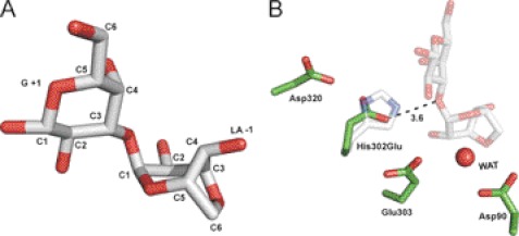
FIGURE 5.
Distortion of substrate and features of catalytic residues. A, the neoagarobiose molecule shows distortion of the 3,6-anhydro-l-galactose into the B1,4 conformation. B, the catalytic residues in the mutant H302E are shown in green and are superposed with the E303Q mutant in complex with neoagarobiose (faint gray). The H302E mutant is inactive likely due to the longer distance between its carboxyl group and the glycosidic bond (3.6 Å) compared with the native His302 residue (2.7 Å). WAT, water.

