FIGURE 3.
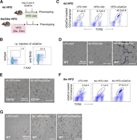
αGalCer challenge activates NKT cells in adipose tissue. A, schematic diagram for three gain-of-function models with 4-day (d) and 8- and 24-week HFD feeding. 6-Week-old mice that have been placed on HFD for 4 days and 8 and 24 weeks were injected intraperitoneally with αGalCer or vehicle (veh) on day 0 and 2 prior to GTT on day 4 and sacrificed for tissues on day 5. B, BrdU labeling of CD1d-tetramer-positive NKT cells in adipose tissue following αGalCer challenge. 7-AAD, 7-aminoactinomycin D. C and F, flow analysis of NKT cells in adipose tissue of mice on 4-day (C) or 8-week (F) HFD. Number refers to the percentage of NKT cells in total CD45+ lymphocytes in stromal vascular cells of adipose tissue. D and E, H&E section of adipose tissue of WT (D) and CD1d−/− (E) mice of 4-day HFD. G, H&E section of adipose tissue of WT mice of 8-week HFD.

