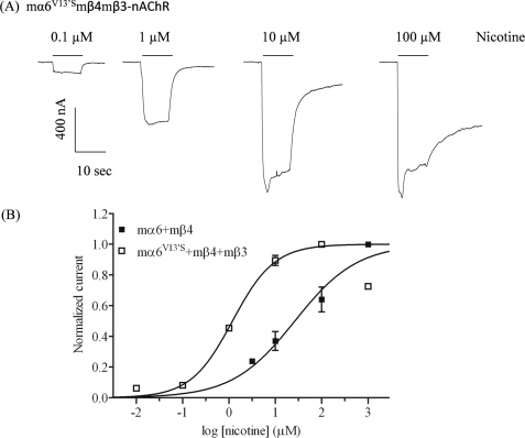
FIGURE 2.
Functional properties of mα6*-nAChR. A, representative traces are shown for inward currents in oocytes held at −70 mV, responding to application at the indicated concentrations of nicotine (shown with the duration of drug exposure as black bars above the traces), and expressing nAChR mα6V13′S, mβ4, and mβ3 subunits. B, results for these and other studies averaged across experiments were used to produce concentration-response curves (ordinate, mean normalized current ± S.E.; abscissa, ligand concentration in log μm) for inward current responses to nicotine as indicated for oocytes expressing nAChR mα6 and mβ4 subunits (■) or mα6V13′S and mβ4 and mβ3 subunits (□), where current amplitudes are represented as a fraction of the peak inward current amplitude in response to the most efficacious concentration of nicotine. Much higher levels of evoked currents are evident for functional nAChR containing mα6V13′S, mβ4, and mβ3 subunits when compared with receptors lacking mα6V13′S subunits. See Table 1 for parameters.

