FIGURE 2.
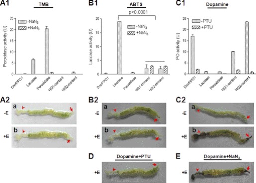
Comparison of the enzyme activity of three oxidases and gut staining. DmrPPO1 was activated by ethanol (E) to have PO activity (16). A1 and A2, peroxidase activity detection. Peroxidase (1.67 ng) and laccase (3 μg) but not DmrPPO1 (1.25 μg) oxidized TMB. No peroxidase activity was detected in HG1 or HG2 content (A1) or in different parts of gut (A2). B1 and B2, laccase activity detection. DmrPPO1 and peroxidase only minimally oxidized ABTS. Some activities were detected in the HG1 and HG2 contents, but they were not inhibited by NaN3 (B1). The real laccase activity was significantly inhibited by NaN3. No laccase activity was detected in the gut (B2). C1 and C2, PO activity detection. Laccase and peroxidase minimally oxidized dopamine. HG1 and HG2 content had obviously high PO activity (C1). The foregut and hindgut stained black only if ethanol was used for activation (C2-b was imaged at 30 min). D and E, effects of laccase and peroxidase inhibitor NaN3 and PO inhibitor PTU on gut staining. PTU significantly inhibited melanization in the foregut and hindgut (D), whereas NaN3 did not (E). Columns represent the mean of individual measurements ± S.E. (n = 3). Significant differences were calculated with an unpaired t test program.

