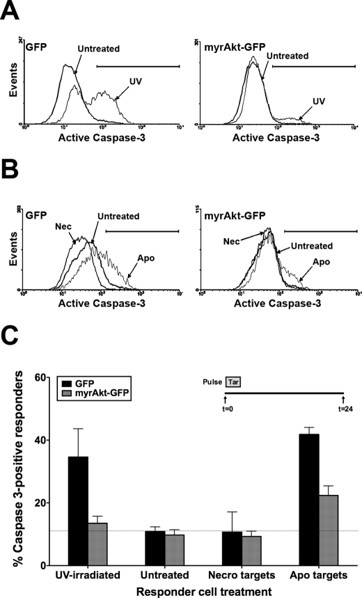
FIGURE 9.
Infection of BU.MPT responders with a constitutively active Akt construct partially prevents loss of viability following exposure to apoptotic targets. BU.MPT responder cells were transiently infected with retroviral constructs containing either GFP alone (GFP) or GFP plus constitutively active myrAkt (myrAkt-GFP). A, infected BU.MPT responders were left untreated (Untreated) or induced to undergo apoptosis by exposure to UV irradiation (UV). Shown is a representative cytofluorimetric analysis for activated caspase-3 24 h after UV irradiation. B, serum-starved infected BU.MPT responders were left untreated (Untreated) or exposed to apoptotic (Apo) or necrotic (Nec) targets at a target/responder cell ratio of 10:1 for 30 min. The source of apoptotic targets was staurosporine-treated DO11.10 cells. Induction of apoptosis in BU.MPT responders was assessed 24 h after exposure to targets by cytofluorimetric analysis of permeabilized responders for activated caspase-3. C, the graph depicts the mean and S.E. from three separate cytofluorimetric analyses of the percentage of BU.MPT responder cells positive for activated caspase-3. In all cases, cytofluorimetric analysis was limited to infected BU.MPT responders whose GFP fluorescence was >1000× that of uninfected cells. The dotted line represents the percentage of caspase 3-positive responders in untreated BU.MPT responders infected with GFP alone. p < 0.05, UV-irradiated and apoptotic targets versus untreated for GFP-infected responders; p < 0.05, UV-irradiated and apoptotic targets, GFP- versus myrAkt-GFP-infected responders; p < 0.01, apoptotic targets versus untreated for myrAkt-GFP-infected responders; p = not significant, UV-irradiated versus untreated for myrAkt-GFP-infected responders. Tar, target(s); Necro, necrotic. Error bars (C) denote S.E.

