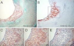
FIGURE 7.
Presence of APLP2 and IαIHC2 within macrophage-rich regions of atherosclerotic lesions. Shown is a section of human aorta stained with Movat's pentachrome (A) to identify cells (pink), elastin (black), glycosaminoglycans (blue), and collagen (yellow); HAM-56 to identify macrophages (B); anti-APLP2 (C); anti-IαIHC2 (D); or anti-serglycin (E).

