FIGURE 6.
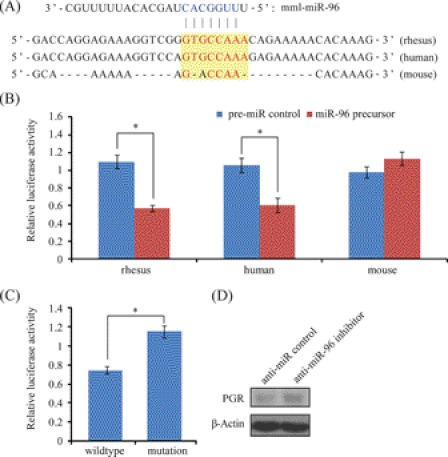
MiR-96 regulation on PGR. A, sequences of predicted miR-96 binding site in the 3′-UTR of PGR. Sequence conservation was compared among rhesus monkey, human, and mouse. The seed sequence of miR-96 was colored in blue and the seed-complementary sequence in the 3′-UTR of PGR is indicated in red. B, target validation by luciferase assay in three different species. The 3′-UTR fragments derived from rhesus monkey, human, and mouse were cloned and subjected to luciferase assay in rhesus LLC-MK2 cells, human ECC-1 cells and mouse TM3 cells, respectively. C, point mutation analysis of miR-96 seed binding sequence. The seed binding site (GTGCCAAA) was mutated into GACCAACA (to mimic the seed binding sequence in mouse). Cells were co-transfected with miR-96 precursor. D, Western blot analysis of PGR-A protein expression after a transfection of anti-miR-96 inhibitor or negative control in rhesus LLC-MK2 cells.

