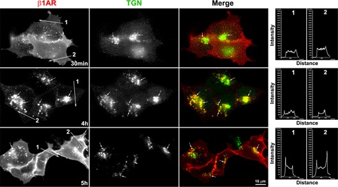
FIGURE 5.
Lysosomal inhibition induces the accumulation of endocytosed β1AR in the TGN. Surface-labeled HA-β1AR cells were incubated with ISO and chloroquine, fixed at various time points, and immunostained for TGN-46. Surface-labeled HA-β1AR and TGN-46 were visualized with donkey anti-rabbit Alexa 594 (red) and anti-sheep Alexa 488 (green), respectively. Co-localization of HA-β1AR with TGN-46 (arrows) is shown in yellow in the merged images. Nuclei were detected with DAPI (blue). Note: whereas the majority of endocytosed HA-β1AR co-localizes with the TGN-46 at 4 h, less HA-β1AR co-localizes with the TGN-46 but more HA-β1AR appears at the plasma membrane at 5 h. The right panels show the pixel intensity profiles of red channels (β1AR staining) along the lines across the plasma membrane. Results are representative of 120 cells from 3 independent experiments.

