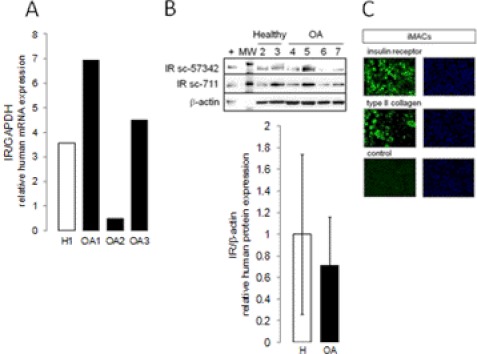
FIGURE 1.
IR expression in articular chondrocytes. A, primary cultures of chondrocytes from healthy (H1 to H3) or osteoarthritic (OA1 to OA7) knees were starved. IR mRNA expression was assessed by real-time RT-PCR. B, IR protein expression was analyzed using immunoblotting with two specific antibodies. Sc-711 (1/200) is a widely used IR antibody; sc-57342 (1/200) is a monoclonal antibody not cross-reacting with IGF-1R. Densitometric signals of IR were normalized to β-actin and expressed as arbitrary units. MW, molecular weight. C, primary cultures of iMACs were starved, and IR protein expression was analyzed using immunocytofluorescence using sc-711. Type II collagen expression was used as positive control of cell differentiation. Specificity of staining was confirmed by omission of primary antibody. Micrographs are representative of three independent experiments performed in triplicate (size bars = 20 μm).

