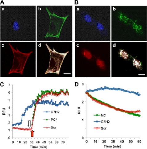
FIGURE 1.
Binding of C7H2 to actin cytoskeleton and actin polymerization. A, co-localization of phalloidin and C7H2 in melanoma cells. B16F10-Nex2 tumor cells were treated with biotinylated C7H2, fixed with paraformaldehyde, and permeabilized with Triton X-100. Cells were stained with DAPI (a), streptavidin-FITC (b), and phalloidin-rhodamine (c) and were examined by confocal microscopy; d, merge, showing co-localization. Scale bar, 20 μm. B, co-localization of DNase I and C7H2 in melanoma cells. B16F10-Nex2 tumor cells were treated with biotinylated C7H2, fixed, and permeabilized as in A. Cells were stained with DAPI (a), streptavidin-FITC (b), and DNase I-Alexa Fluor 594 (c) and were examined by confocal microscopy; d, merge, showing co-localization. Scale bar, 20 μm. C, pyrene G-actin (0.4 mg/ml) added to a 96-well plate. C7H2 at 200 μm (●, blue) and the scramble peptide Scr-C7H2 (△, red) were added, and the fluorescence at 410 nm was measured in kinetic mode. At the end of 30 min (open arrow) or at the same time without previous incubation with peptide (red arrow), the ATP-containing polymerization buffer was added as a positive control (PC*, green). D, pyrene F-actin from stock incubated for 1 h diluted to 0.2 mg/ml and added to a 96-well plate. C7H2 at 200 μm (●, blue) and the scramble peptide Scr-C7H2 (△, red) were added, and the fluorescence at 410 nm in kinetic mode was measured. Negative control, NC (♦, green), incubation with the G-buffer.

