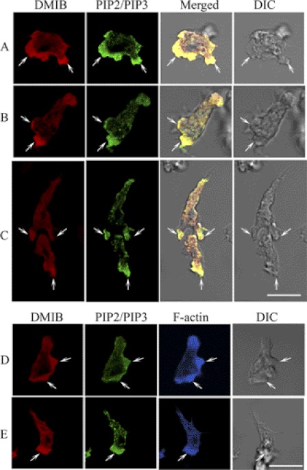
FIGURE 1.
Colocalization of endogenous DMIB and PIP2/PIP3 in fixed AX3 cells. AX3 cells were fixed and endogenous DMIB, and PIP2/PIP3 were visualized with antibodies against DMIB (red) and PIP2/PIP3 (green). F-actin was stained with phalloidin Alexa Fluor 633 (blue). In nonpolarized cells, DMIB colocalizes with PIP2/PIP3 in random cell protrusions (A). In polarized cells, DMIB and PIP2/PIP3 colocalize at the cell front (B). In chemotaxing cells, DMIB and PIP2/PIP3 colocalize at the front of the leading cell and in the engulfing mouth of the following cell (C). Actin colocalizes with DMIB and PIP2/PIP3 at random cell protrusions (D) and at the front of polarized cells (E). Arrows mark the sites of colocalization of DMIB with PIP2/PIP3 (A–C) and with F-actin (D and E). Scale bars, 10 μm. DIC, differential interference control microscopy.

