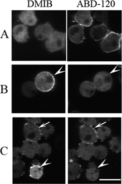
FIGURE 10.
Relocation of DMIB to plasma membrane in cells treated with LatA. AX3 cells cotransfected with DMIB and F-actin probe ABD-120 were starved for 4 h and treated with 7.5 μm LatA. A, in cells before treatment DMIB is mostly diffused and cortical actin is present. B and C, after 20-min exposure to LatA, cortical F-actin is absent, and DMIB reappears on the plasma membrane, alone (arrowheads) or accompanied by F-actin patches (arrows). Scale bar, 10 μm.

