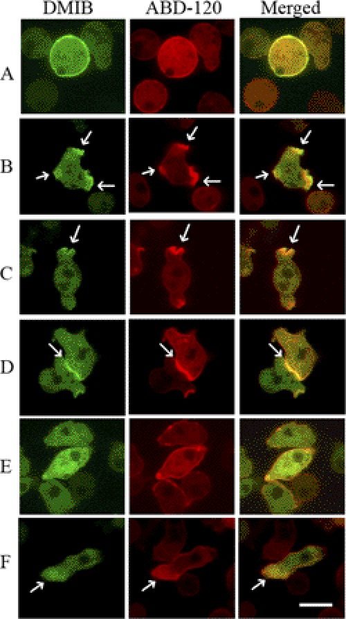
FIGURE 2.
Localization of expressed GFP-DMIB in live cells. AX3 cells were cotransfected with DMIB and ABD-120 fused to GFP (green) and RFP (red), respectively. Live cells images are shown. DMIB is localized uniformly on the plasma membrane of freshly plated cells (A), in pseudopods (B), cups (C), and at cell-cell contacts (D) of randomly moving cells. DMIB is mostly diffuse in cells starved for about 4 h (E) and is localized to the cell front in elongated polarized cells (F). In all cases except E, DMIB colocalizes with F-actin. Arrows mark sites of DMIB and F-actin colocalization. Scale bar, 10 μm. See also supplemental Movie S1.

