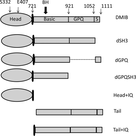
FIGURE 3.
Schematic representation of DMIB and its mutants expressed in Dictyostelium cells. The major regions that were deleted are labeled (S = SH3). The positions of residues mutated within the head and the position of BH site are indicated. The point mutations in the head in full-length DMIB were: S332A (DMIB-S332A), which results in motor-dead myosin, and E407K (DMIB-E407K), which results in severely reduced binding to F-actin. Mutations of the BH sites in full-length DMIB, dGPQSH3, and the tail were: deletion of entire BH site (dBH), substitution of 5 basic residues within the BH site with Ala (BH-Ala), and point mutation I810D. See supplemental Fig. S2 for more detailed description of DMIB-I810D.

