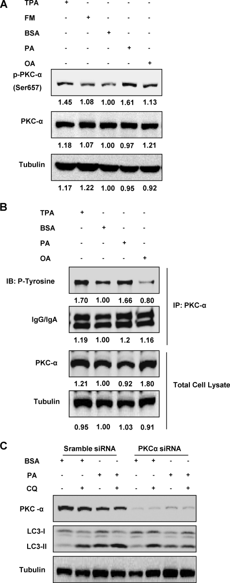
FIGURE 6.
Activation of PKC-α is required for PA-induced autophagy. A, MEFs were treated with BSA control, PA (0.25 mm), or OA (0.25 mm) for 4 h, and activation status of PKC-α was observed using immunoblotting. Cells treated with TPA (100 nm) for 20 min were used as a positive control. The respective lane intensity was quantified using Kodak Imaging Software as a -fold change to the BSA control treatment. B, MEFs were treated with BSA control, PA (0.25 mm), or OA (0.25 mm) for 4 h, and PKC-α was then immunoprecipitated (IP) and blotted for levels of phosphorylated tyrosine as another indicator of PKC-α activation. TPA (100 nm) was added to the cells for 20 min to act as a positive control. The respective lane intensity was quantified using Kodak Imaging Software as a -fold change to the BSA control treatment. C, PKC-α in MEFs was knocked down with PKC-α siRNA, whereas the control cells were knocked down with scramble siRNA as described. The cells were then treated with either BSA control or PA (0.25 mm) for 4 h with or without the presence of CQ (10 μm).

