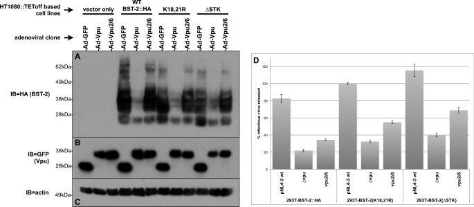
FIGURE 5.
BST-2s cytoplasmically exposed serine and threonine residues are not required for down-regulation of total levels of BST-2 by Vpu nor are they required for Vpu to promote viral release. A, HT1080::TEToff-based cell lines expressing vector (control), WT, ΔK (K18R,K21R), and ΔSTK BST-2 proteins were infected with the same Vpu-expressing adenoviruses described above in Fig. 2, B–D. An immunoblot (IB) of the resulting cell lysates was successively probed with HA (panel A, to detect BST-2::HA), GFP (panel B, to detect Vpu fusions), and actin antibodies (panel C, loading control). D, shown are TZM-bl viral egress assay data for supernatants collected from 293T-based cells lines (expressing the same BST-2 mutants shown in the immunoblots) transfected with the indicated pNL4–3-based proviral clones. Egress data are expressed as a percentage of viral release from the control 293T-based cell line that does not express BST-2.

