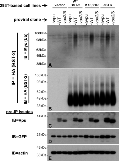
FIGURE 6.
The BST-2 cytoplasmically exposed serine and threonine residues are not required for the specific ubiquitination of BST-2 by Vpu. BST-2 ubiquitination assays were performed essentially as described for Fig. 2, with the exception that all samples were treated with CMA. Immunoblots (IB) of HA-bead IPs from each sample were first probed for Ub (panel A, Myc) then stripped and reprobed for HA (panel B, to detect BST-2::HA). Samples of the pre-IP lysates (bottom three panels) were sequentially probed for Vpu (C), GFP (D, transfection control), and finally actin (E, loading control).

