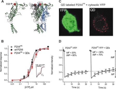
FIGURE 2.
HA tag at Gln-78 in P2X4HA receptors is surface-exposed and can be labeled with QDs without altering receptor function or macroscopic mobility. A, models show a single P2X4 subunit (left) and a trimeric P2X4 receptor (right) with the HA tag insertion site shown schematically as a red ball (at Gln-78) in the extracellular domain. B, normalized concentration-effect curves for ATP at WT P2X4, P2X4HA, and P2X4HA receptors transfected into microglia and labeled with 100 pm QDs to ensure that most receptors were bound to QDs (n = 8, 6, and 6). The inset shows normalized representative 100 μm ATP-evoked currents from the three concentration-effect curves. C, representative image of a HEK cell expressing P2X4HA receptors and cytosolic YFP. The QD image was obtained using a high amount of QDs (100 pm) to ensure most surface receptors were labeled. D, FRAP curves for P2X4HA-YFP receptors with (n = 20) and without QD labeling (n = 12). There was no decrease in the mobile fraction (MF) from FRAP recovery curves. If anything, there was a small trend for the mobile fraction to be greater after QD labeling (36 ± 3 versus 22 ± 3% for P2X4HA-YFP and P2X4HA-YFP + QD receptors, respectively; p < 0.01 with an unpaired Student's t test). Overall, these data indicate that QD labeling did not decrease P2X4 receptor mobility.

