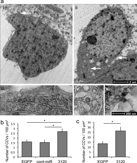
FIGURE 6.
miR-3120 prevents uncoating of clathrin-coated vesicles. a, transmission electron microscopy image of EGFP transduced cell bodies (images i and ii) identified by the presence of a nucleus and surrounding cytoplasm (1,900 times end magnification). White arrow in image i denotes the pit shown in the (13,000 times) high magnification image (image iii), whereas the arrows in image ii show (13,000 times) high power images (images iv and v) of clathrin-coated vesicles. Image iii also shows ribosomes on the endoplasmic reticulum. b, clathrin-coated vesicle (CCV) counts in cortical neurons transduced with miR-3120 when compared with those transduced with EGFP and the miRNA control (Cont-miR). A similar increase in clathrin-coated vesicle number was seen in hippocampal neurons transduced with miR-3120. A detailed description of the procedures for counting clathrin-coated vesicles is given under “Results.” For both hippocampal and cortical neuronal cultures, a minimum total of 17 cells was analyzed in three individual experiments. Values are means ± S.E. Statistical analyses were conducted by analysis of variance and post hoc Bonferroni's test * = p > 0.01.

