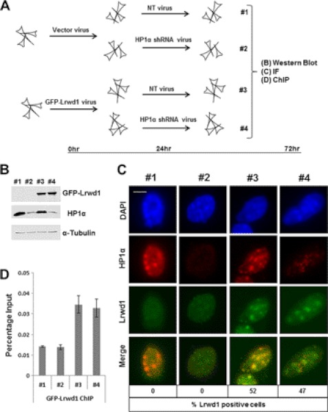
FIGURE 6.
Pericentric heterochromatin localization of Lrwd1 is independent of HP1α. A, diagram shows the experimental procedures. B, Western blotting analysis of expression of GFP-Lrwd1 and HP1α in four samples from A, C, and D is shown. C, localization of Lrwd1 is unaffected in HP1α-depleted cells compared with nontargeting cells. Cells with Lrwd1 localization at pericentric heterochromatin were counted (n = 100) in three independent experiments. Scale bar, 5 μm. D, depletion of HP1α does not affect Lrwd1 binding to pericentric heterochromatin. ChIP assays using antibodies against GFP-Lrwd1 were performed, and ChIP DNA was analyzed by real-time PCR using PCR primers amplifying major satellite repeat.

