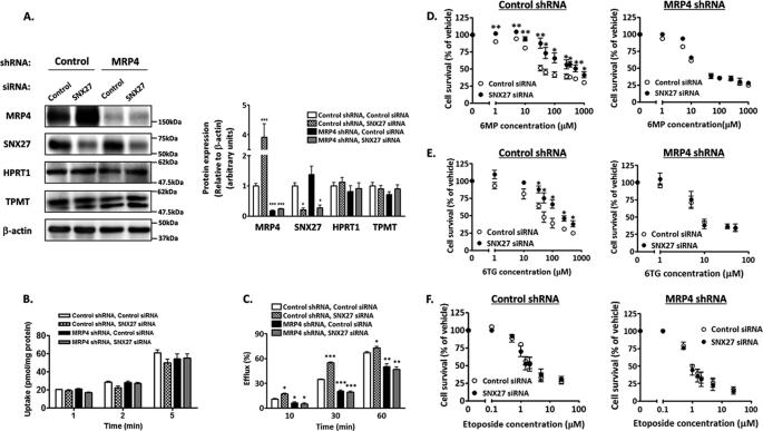
FIGURE 4.
Effect of SNX27 depletion on resistance to 6MP and 6TG. HEK-MRP4 shRNA or HEK-control shRNA cells were transfected with SNX27 siRNA or control siRNA. After 24 h (D–F) or 48 h (A–C) of transfection, these cells were studied. A, expression of MRP4, SNX27, HPRT1, and thiopurine methyltransferase. Left, cell lysates were prepared and subjected to Western blot analysis. A representative result from four independent experiments is shown. Right, Image Gauge software was used to quantify the ratios of band intensities of each protein to that of β-actin. Each bar represents the mean ± S.E. (error bars) of four independent experiments. *, p < 0.05; ***, p < 0.001 compared with control siRNA-transfected HEK-control shRNA cells. B, time profiles of uptake of [14C]6MP. Cells were incubated at 37 °C in KH buffer containing 5 μm [14C]6MP, and its time-dependent uptake was determined. Each bar represents the mean ± S.E. of three independent experiments. C, time profiles of efflux of [14C]6MP. Cells were incubated for 2 h as described in B, and the time-dependent efflux was determined. Each bar represents the mean ± S.E. of three independent experiments. ***, p < 0.001; **, p < 0.01; *, p < 0.05 compared with control siRNA-transfected HEK-control shRNA cells. D–F, sensitivity to 6MP, 6TG, and etoposide. Cells were incubated for 96 h in medium containing 6MP (C), 6TG (D), or etoposide (E) at the concentration indicated. Drug sensitivity was determined as described under “Experimental Procedures.” Each point represents the mean ± S.E. from three independent experiments in triplicate. **, p < 0.01; *, p < 0.05.

