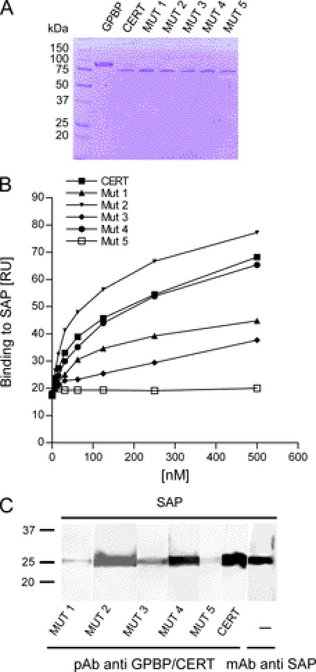
FIGURE 11.
Binding of CERT and CERT mutants to SAP. A, Coomassie staining of immunopurified wild type and mutant 1–5 CERT proteins (2 μg of each sample were loaded per lane). B, 2-fold serial dilutions (3.9–500 nm) of wild type and mutant 1–5 CERT proteins in 25 mm HEPES, pH 7.4, 150 mm NaCl with 0.01% Tween 20 were tested for binding on immobilized SAP by SPR. At each concentration, the highest binding signal was measured. C, SAP was separated on a 12% SDS-polyacrylamide gel, electroblotted to nitrocellulose, and prepared for far Western analyses as described in the legend to Fig. 4A. The membrane with renatured SAP was cut into strips and probed with CERT mutants (lanes 1–5), WT (lane 6), or mAb anti-SAP (lane 7). Membrane strips incubated with CERT proteins were detected with anti-GPBP/CERT 300–350.

