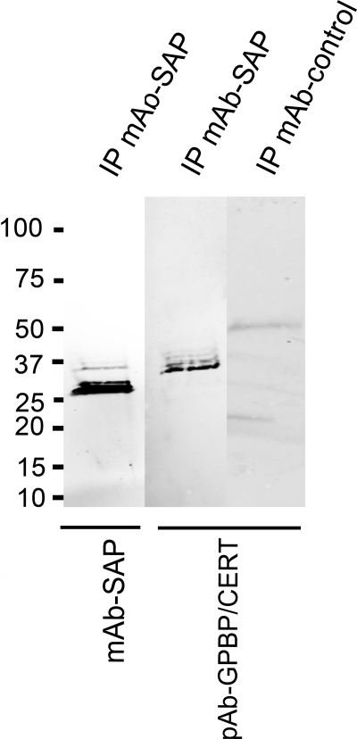
FIGURE 8.
SAP-GPBP complexes in plasma. SAP was immunoprecipitated with mouse mAb anti-SAP (clone 4E8, Sigma), followed by immunoblotting with the same antibody to detect immunoprecipitated SAP (first lane) and a polyclonal antibody against GPBP/CERT 1–50 to detect GPBPs (second lane). The control using an isotype control mAb (mouse monoclonal anti-syntaxin 6, clone 3D10) for immunoprecipitation (IP) was negative when detected with the same polyclonal antibody against GPBP (third lane). GPBP is efficiently co-precipitated with SAP in a band of ∼37 kDa, suggesting that a part of the GPBP molecule corresponding to the N-terminal domains of the protein interacts with SAP. Results shown are representative of five experiments.

