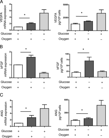
FIGURE 1.
Glucose deprivation induces the expression of angiogenic mediators. A, HREC were cultured in glucose-deprived medium or in standard medium with or without hypoxia for 24 h. Left: VEGFA transcript levels were measured by qRT-PCR and are presented as the mean ± S.E. relative to levels after 24 h of culture with standard conditions in four independent experiments. Right: secretion of VEGFA in the medium was quantified by ELISA. The concentration is presented as the mean ± S.E. of three independent experiments. *, p < 0.05. B, HREC were cultured in glucose-deprived medium or in standard medium with or without hypoxia for 24 h. Left: bFGF transcript levels were measured by qRT-PCR and are presented as the mean ± S.E. relative to levels after 24 h of culture with standard conditions in four independent experiments. Right: secretion of bFGF in the medium was quantified by ELISA. The concentration is presented as the mean ± S.E. of three independent experiments. *, p < 0.05. C, HREC were cultured in glucose-deprived medium or in standard medium with or without hypoxia for 24 h. Left: ANG transcript levels were measured by qRT-PCR and are presented as the mean ± S.E. relative to levels after 24 h of culture with standard conditions in four independent experiments. Right: secretion of ANG in the medium was quantified by ELISA. The concentration is presented as the mean ± S.E. of three independent experiments. *, p < 0.05.

