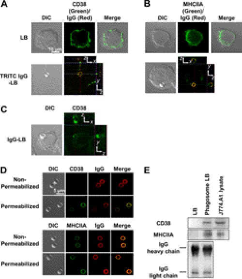
FIGURE 2.
Two or three-dimensional fluorescence images of CD38 and myosin heavy chain IIA in J774A.1 murine macrophages. CD38 and MHCIIA internalization via FcγR-mediated phagocytosis was visualized through immunohistochemistry with a primary FITC-conjugated rat anti-mouse CD38 or a rabbit polyclonal MYH9 antibody, respectively, as described under “Experimental Procedures.” A, CD38 (green) was found to be situated along the plasma membrane in its resting state (control). Initiation of FcγR-mediated phagocytosis with TRITC IgG-opsonized latex beads (LB, red) resulted in CD38 internalization where it co-localized with the phagosome. DIC, differential interference contrast. B, MHCIIA (green) was also found to be situated along the plasma membrane in its resting state (control). Initiation of FcγR-mediated phagocytosis with TRITC IgG-opsonized latex beads (red) resulted in MHCIIA internalization, where it also colocalized with the phagosome. C, CD38 (green) also internalizes independently of the phagosome after initiating FcγR-mediated phagocytosis with IgG-opsonized latex beads. The blue line shows the position of the phagosome on the z axis. The x (green line), y (red line) image is the confocal z axis slice corresponding to the position of the blue line (i.e. through the center of the phagosome). The phagosome is stably positioned within the cell. D, shown are co-localized images of CD38 or MHCIIA with phagocytosed-latex bead. Latex bead-containing phagosomes were isolated as described under “Experimental Procedures” and stained with anti-CD38 and anti-MHCIIA antibodies with or without permeabilization. E, shown is a Western blot of CD38 and IgG with biotinylated anti-CD38 antibody and anti-mouse IgG antibody on latex beads (LB), latex bead-containing phagosome (phagosome LB), and whole lysate of J774.A1 cells.

