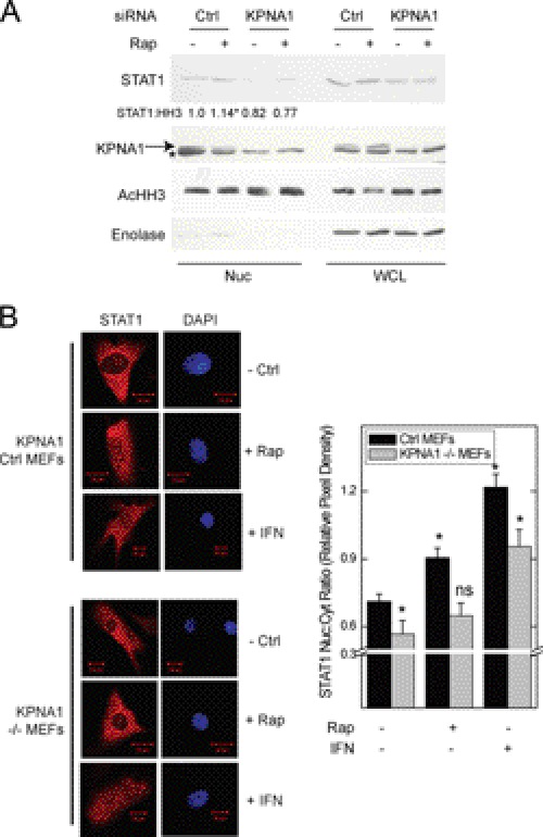
FIGURE 4.
KPNA1 is required for the enhancing effect of rapamycin on STAT1 nuclear content. A, A549 cells were transfected with control (Ctrl) siRNAs or those targeting KPNA1 for 72 h before incubation for 1 h with fresh medium without or with 50 ng/ml rapamycin (Rap) preparation of nuclear (Nuc) or whole cell (WCL) lysates and detection of the indicated proteins by Western blot. The arrow indicates KPNA1, and the asterisk indicates another non-KPNA1 reactive band. Gels are representative of four individual experiments. Nuclear STAT1 protein levels are quantified as fold change in band density relative to control = 1 (means ± S.E.). *, p < 0.05 versus control-transfected cells without rapamycin). B, control or KPNA1-deficient (−/−) MEFs were incubated without or with 50 ng/ml rapamycin or IFN-γ, 100 units/ml, for 0 (− Rap) or 1 h (+ Rap). Endogenous STAT1 (red) was detected by indirect immunofluorescence confocal microscopy. The slides were mounted with solution containing the nuclear marker DAPI (navy blue). Shown to the right of representative images are summarized data (mean nuclear to cytoplasmic pixel density ratio ± S.E., n = 3–5 cells/experiment) representative of five independent experiments. *, p < 0.05 versus control; ns, not significant by Student's t test.

