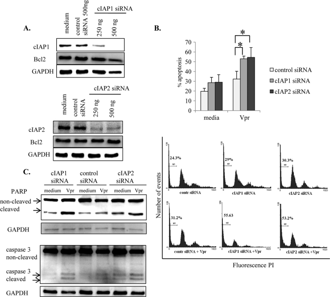
FIGURE 10.
IAPs protect macrophages against Vpr-induced apoptosis. A, after differentiation, THP1-MACs (5 × 10⁵/ml) were transfected with cIAP1, cIAP2, or control siRNA as described under “Experimental Procedures.” Following transfection, cells were collected after 24 h for evaluation of protein knockdown. Total cell proteins were subjected to Western blotting, and the membranes were probed with antibodies specific for cIAP1, cIAP2, and Bcl2 to ensure siRNA specificity. B and C, on the second day after transfection, cells were treated with Vpr (2.5 μm) for another 24 h and stained with PI for apoptosis measurement (B) or subjected to Western blotting for PARP and caspase-3 cleavage (C). Only 500 ng of IAP siRNA and control siRNA were used for these experiments. The bar graph in the top panel of B shows the mean percentage of apoptosis ± S.D. (error bars) of four separate experiments. *, p < 0.05. Histograms in the bottom panel of B show one representative experiment indicating the percentage of cells with subdiploid DNA content.

