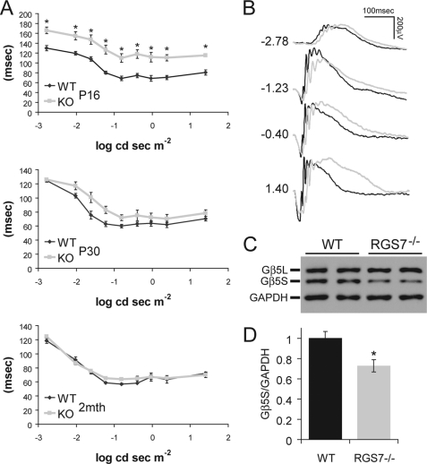
FIGURE 2.
RGS7−/− mice exhibit an age-dependent delayed ERG b-wave implicit time as well as loss of Gβ5S protein in the retina. A, ERG b-wave implicit time, measured as the time difference between flash onset and the time b-wave is at its peak, was significantly delayed (*, p < 0.01) after eye opening at P16 in RGS7−/− mice (KO, n = 8) compared with WT littermate controls (n = 8). At P30, the KO (n = 6) implicit time delay was improved compared with WT (n = 6), showing a slight trend, but this delay was no longer statistically significant. At 2 months of age (2mth), the b-wave implicit time of KO (n = 8) mice is indistinguishable from that of WT animals (n = 6). Error bars are S.E. B, representative ERG traces from WT (black) and KO (gray) animals at P16 are shown with flash intensity indicated at left in unit of log cd s m−2. C and D, Gβ5S protein level in the retina was decreased in adult RGS7−/− mouse at P30. C, representative immunoblot shows the level of Gβ5S from two WT and two RGS7−/− mice retinal protein extracts (20 μg) using anti-Gβ5 (CT215, 1:2,000) and anti-GAPDH (1:50,000) antibodies. D, quantification of Gβ5S protein level, normalized to GAPDH signals, shows a significant 27 ± 7% reduction in the RGS7−/− (n = 6) versus WT (n = 4) mice (*, p < 0.01). Error bars are S.E.

