FIGURE 4.
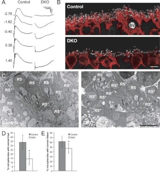
RGS7 and RGS11 dKO mice have no ERG b-waves and contain ultrastructural defects in the retinal OPL. A, representative scotopic ERG responses from 2-month-old WT control and 711dKO (DKO) mice with flash intensity indicated at left in units of log cd s m−2. B, immunohistochemical staining of control and DKO retinal sections for CtBP2 (1:2,000, white) and PKCα (1:100,000, red) demonstrating reduction in rod bipolar cell dendritic arborization. BV, blood vessel. Scale bar, 5 μm. C, representative transmission electron microscopic images of the OPL from control and DKO mice. Scale bar, 2 μm. RS, rod spherule. Asterisk denotes abnormal structure. D and E, percentage of rod spherules with normal synaptic triads (D) and ribbons (E) in the DKO (n = 4, 80 images) versus control (n = 3, 65 images) retinas (*, p < 0.01). Error bars are S.D.

