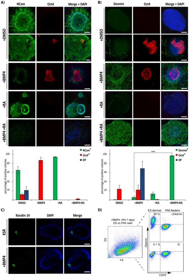
Figure 1. Effect of BMP4 on differentiation of ES cells cultured on PA6 feeder cells.
ES cells were induced to differentiate on PA6 cells as indicated and analysed by immunofluorescence microscopy (A, B and C) or flow cytometry (D). The immunofluorescence images show expression of NCam and Oct4 (A), Desmin and Oct4 (B) or Keratin 14 (C). Quantification of the immunofluorescence experiments from (A) and (B) is shown in the lower bar diagrams. Colonies were counted as single positive for NCam (NCam+), Oct4 (Oct4+) or Double positive (DP) in (A); and single positive for Desmin (Des+), Oct4 (Oct4+) or double positive (DP) in (B). Data are represented as the average ± Standard Error of the Mean (SEM) of 3 independent differentiation experiments (***, p<0.001, n = 3). (D) ES cells were induced to differentiate for 7 days with BMP4 and RA on CSFE-labelled PA6 and Desmin expression was assessed by flow cytometry (right hand dot plots). Side (SS) and forward scatter (FS) profiles are shown in the left hand dot plot. IC: isotype control antibody. Scale bars, 50 µm.

