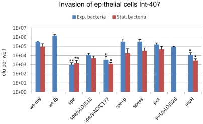
Figure 3. Invasion of epithelial cells.
Int-407 cells were infected with exponential phase M9 cultures (blue bars) or overnight M9 cultures (red bars) of the indicated strains. Non-adherent bacteria were removed and adherent bacteria were enumerated by plating (not shown). For determination of invasion extracellular bacteria were killed by gentamicin and intracellular bacteria were enumerated by plating. The strains tested are: wt; S. Typhimurium 4/74, spe (biosynthesis), pot (transport), invH; SPI1 invasion mutant, spe/pACYC177; spe-mutant with blank complementation plasmid, spe/pLOJ318; spe-mutant complemented with speB (putrescine biosynthesis). pot/pLOJ326; pot-mutant complemented with potCD (spermidine/putrescine uptake). +p and +s denotes that the bacterial cultures have been supplemented with 100 µg ml−1 of putrescine or spermidine, respectively, prior to invasion. The experiments were repeated at least 4 times with similar results and shown is an average of these. Errorbars indicate standard deviations. Significant differences between the wt and the mutants are indicated with aterixs (* P<0.05; ** P<0.001). The P-values were calculated by a one-way ANOVA using Bonferronís post-test.

