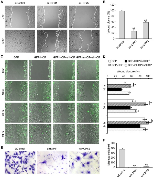
Figure 3. HOP is important for endothelial cell migration.
A. HUVECs transfected with HOP or control siRNAs were scratched, and wound margins were imaged 0 or 19 hours later. B. Experiments were performed as in A, and the extent of wound closure was quantified by measuring the wound area compared with the initial wound area. **, p<0.01 versus control. C. HUVECs transfected with GFP, GFP-HOP, GFP-HOP plus HOP shRNA, or GFP-mHOP plus HOP shRNA were scratched, and wound margins were imaged 0, 10, 20, or 24 hours later. D. Experiments were performed as in C, and the extent of wound closure was quantified. *, p<0.05. E. HUVECs transfected with HOP or control siRNAs for 84 hours were placed in transwell migration chambers containing filters coated uniformly on upper side with matrigel. To stimulate migration, 10% FBS was added to the lower chamber. Transwell chambers were stained with crystal violet and imaged after 12 hours. F. Experiments were performed as in E, and the average number of migrated cells was quantified. **, p<0.01 versus control.

