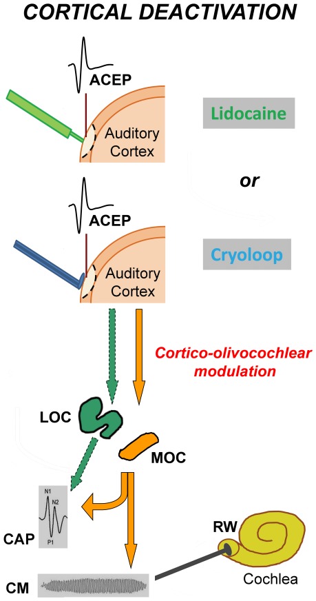
Figure 1. Schematic illustration of experimental procedures.
In all deactivation experiments (n = 15), auditory cortex evoked potentials (ACEP) were recorded from the left auditory cortex, and cochlear potentials (CAP and CM) from the right round window (RW) (contralateral experiments), while in two cases, left cochlear potentials were also obtained (bilateral experiments). Two methods were used in separate experiments to deactivate the auditory cortex: (i) cortical microinjection of lidocaine (n = 10) and (ii) cortical cooling with cryoloops (n = 5). Cortico-olivocochlear effects of auditory cortex deactivation were evaluated by measuring cochlear and auditory neural responses (CM and CAP) before and after cortical deactivation. We propose the presence of two descending pathways from the auditory cortex to the inferior colliculus (not shown in this figure), and from IC to medial and lateral olivocochlear neurons, represented in orange (MOC) and green (LOC) colors respectively. A corticofugal modulation of MOC activity (orange colored arrows) would modify both CAP and CM responses, while a cortical modulation of LOC activity (green colored arrows) would only affect CAP responses.

