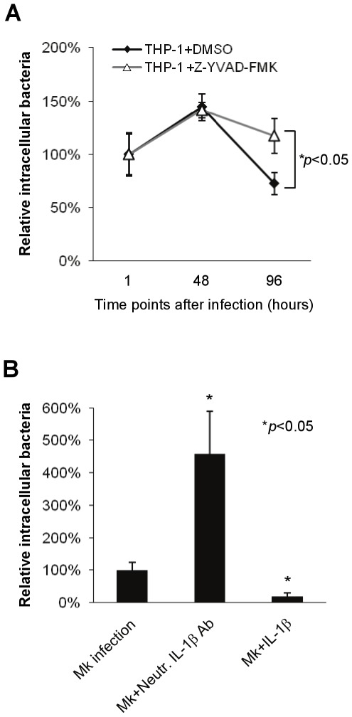
Figure 5. Caspase-1 activation and IL-1β secretion restrict M. kansasii growth.
(A) THP-1 macrophages were infected with M. kansasii at an MOI of 1 for 1 h, before treatment with 50 µM caspase-1 inhibitor (Z-YVAD-FMK) or dimethylsulfoxide (DMSO) alone as control. Cells were then lysed and the intracellular bacterial load was quantified at indicated time points. (B) The M. kansasii-infected macrophages (MOI 1, 1 h) were treated with neutralizing antibodies specific for IL-1βor with exogenous IL-1β for 48 h. The intracellular bacterial CFU was then determined. Results represent the mean ± standard deviations of three independent experiments. Data were analyzed by Student's t test. *p<0.05 compared to untreated infected cells.

